
Current research
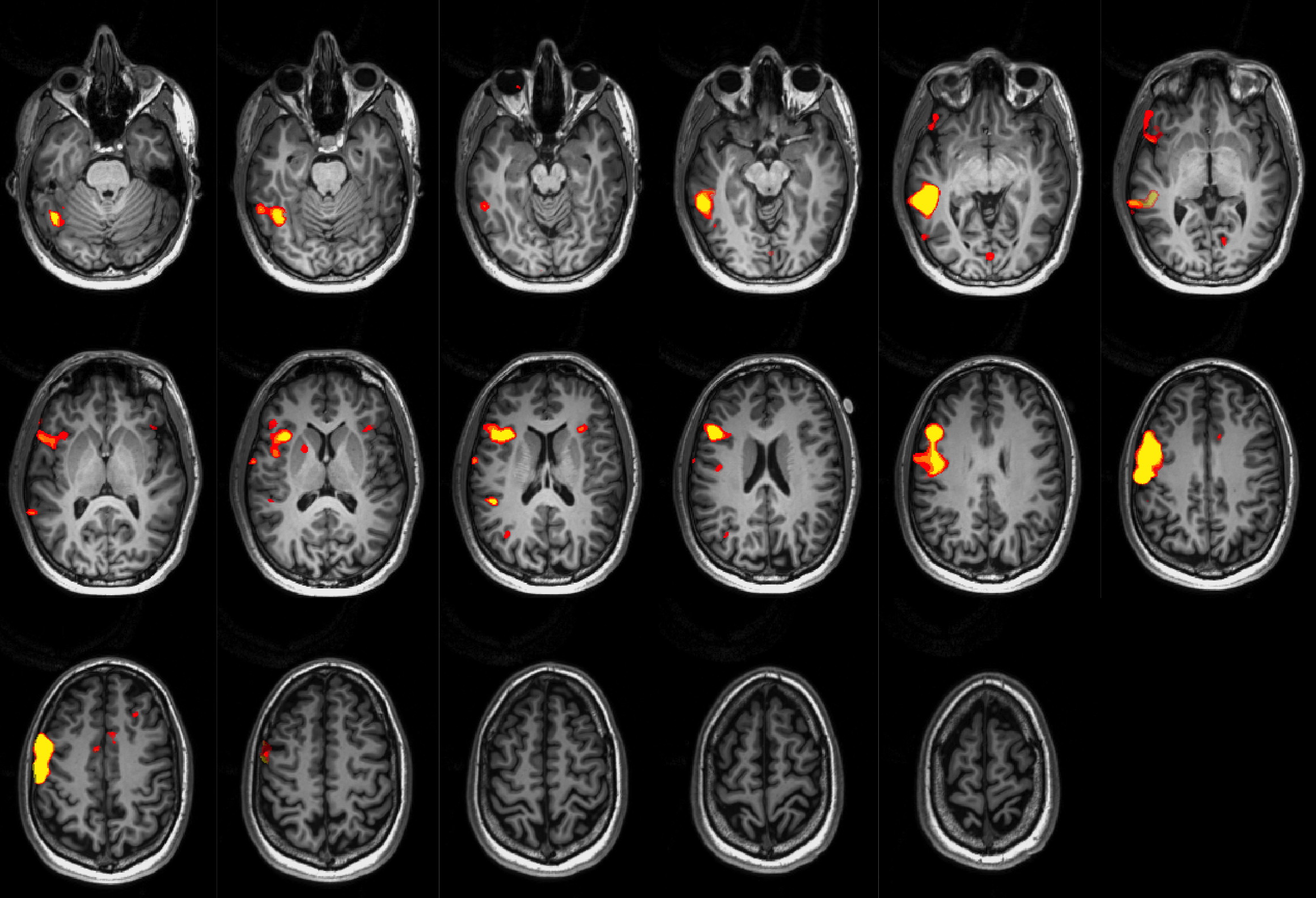
Language fMRI in a patient with right hemisphere dominance. Right of the image is the left of the brain (radiological conventions).
A critical goal of surgical planning is that we can identify all language areas to plan any possible surgery. fMRI is currently validated to identify the language dominant hemisphere, but not to localize specific areas of language cortex. In an early study I showed fMRI could parse different language regions and their interaction in typical children and in children suffering language disorder (impaired fluent reading). In a subsequent set of studies at UCLA we have examined the relationship between Wada test results and fMRI; examined the influence of speaking multiple languages on language system organization; and provided initial evidence that including assessment of grammar in Wada testing may improve the test's utility.
My current work focuses on mapping language in pre surgical planning.
In an initial study (Benjamin et al., 2017) we have studied the ability of fMRI to localize a set of six different language regions. I am studying the validity and reliability of this method for localizing specific language regions, and using this information to improve the ability of the current gold-standard methods–such as cortical stimulation–to map language and improve surgical planning. In a second survey study we sought to understand how epilepsy sites world-wide currently implement and interpret fMRI language maps. Manuscripts based on this work are currently in preparation. This work has been kindly supported by the National Academy of Neuropsychology, Yale Neurology and the Yale Center for Clinical Investigation.